About Our Research
The Clinical Nephrology Section aims to provide and support both inpatient and outpatient comprehensive kidney consultation services and kidney replacement therapy care to any patient enrolled in clinical research protocols at the NIH Clinical Center upon the request of Principal Investigators. The section strives to offer high-quality kidney specialty care to patients with a wide variety of complex kidney diseases. This includes care for patients with hypertension, fluid and electrolyte disorders, acute kidney injury, and chronic kidney disease. Additionally, the section is committed to training physicians in the specialty of nephrology at the residency and fellowship level and providing training in basic, translational and clinical research related to the kidney. Furthermore, the section is dedicated to promoting the study, discovery, and treatment of rare kidney disorders, contributing to the advancement of nephrology. The section is actively involved in conducting studies on the pathogenesis and therapy of glomerular diseases. Currently, there is a particular focus on researching Membranous Nephropathy (MN) and focal segmental glomerulosclerosis (FSGS) aiming to improve understanding and treatment. Through these endeavors, the Clinical Kidney Section commits to improving patient care and advancing kidney disease research.
The Clinical Nephrology Section’s mission is to:
- Provide and support both inpatient and outpatient comprehensive kidney consultation services and kidney replacement therapy care to any patient enrolled in clinical research protocols at the NIH Clinical Center at the request of Principal Investigators.
- Provide high-quality kidney specialty care to patients with a wide variety of complex kidney diseases, including those with hypertension, fluid and electrolyte disorders, acute kidney injury, and chronic kidney disease.
- Provide training in nephology and in research related to kidney disorders.
- Promote the study, discovery, and treatment of rare kidney disorders.
Current Research
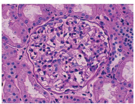
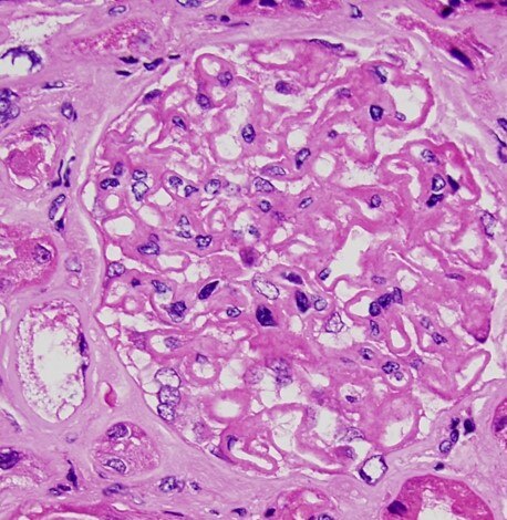
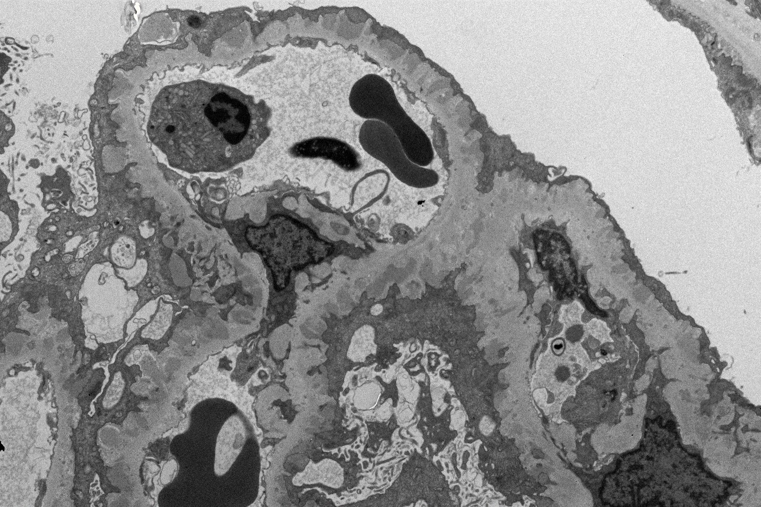
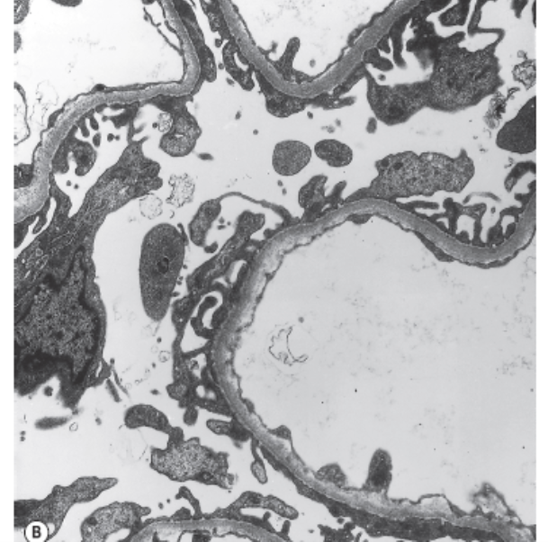
The Clinical Nephrology Section studies various kidney diseases, including membranous nephropathy and focal segmental glomerulosclerosis. The goal of our research is to better understand the evolution and outcomes of these kidney diseases and find treatments that may be more effective and less toxic.
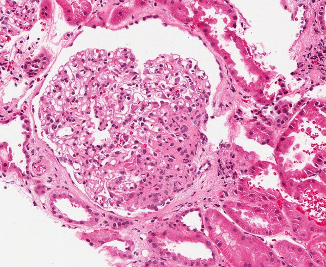
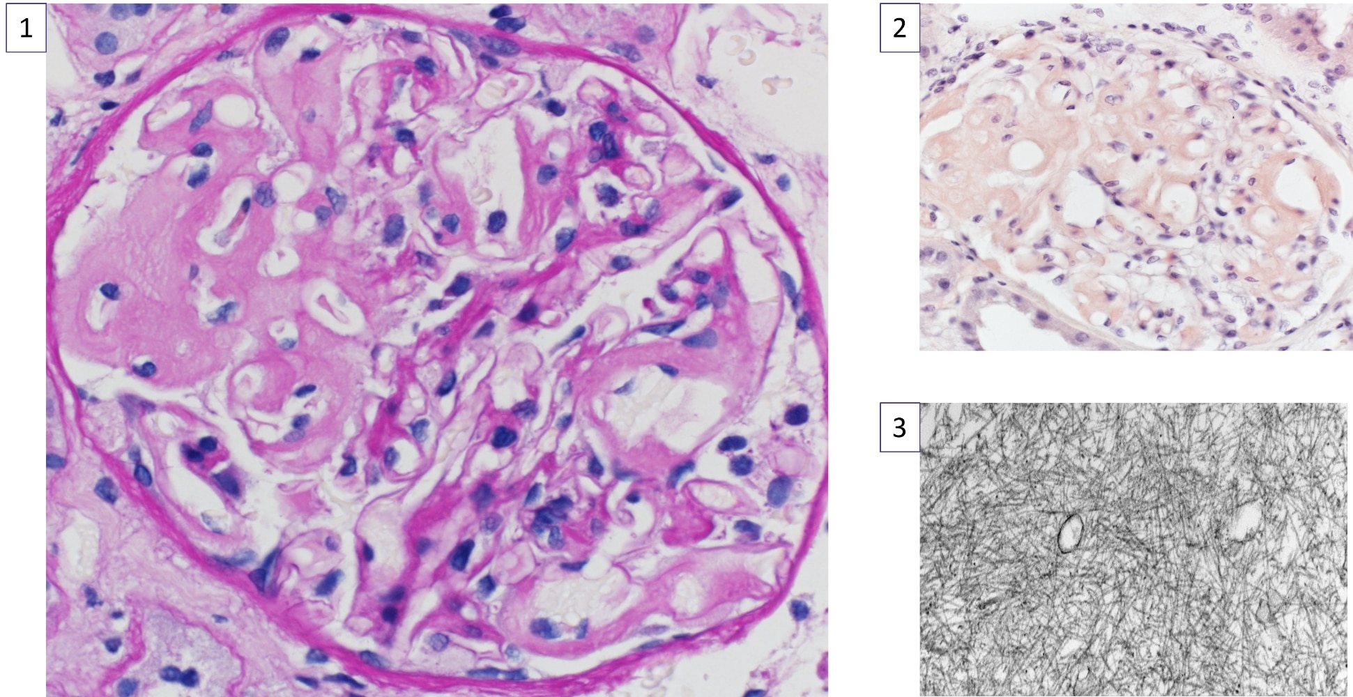
Research Images
![Glomerular capillary loops.](/-/media/Images/labs-branches-sections/kidney-disease-branch/clinical-nephrology-section/research-images/normal-glomerular-architecture_533x410.png)
![Kidney biopsy showing thickening of capillary loops in the glomerulus typical of membranous nephropathy.](/-/media/Images/labs-branches-sections/kidney-disease-branch/clinical-nephrology-section/research-images/kidney-biopsy-membranous-nephropathy_458x470.jpg)
![Dark projections representing immune deposits that form on top of the glomerular basement membrane in the kidney disease membranous nephropathy.](/-/media/Images/labs-branches-sections/kidney-disease-branch/clinical-nephrology-section/research-images/membranous-nephropathy_1551x1035.png)
![Ultrastructure of a normal glomerular capillary wall.](/-/media/Images/labs-branches-sections/kidney-disease-branch/clinical-nephrology-section/research-images/ultrastructure-normal-glomerular-capillary_609x602.png)
![Kidney biopsy showing focal and segmental scarring of glomerulus typical of FSGS.](/-/media/Images/labs-branches-sections/kidney-disease-branch/clinical-nephrology-section/research-images/kidney-biopsy-fsgs_667x545.png)
![A collage of slides of a kidney biopsy. Image 1 shows a single glomerulus from a kidney biopsy. Within the glomerulus, there are deposits that appear acellular and are negative for periodic acid-Schiff (PAS) staining. These deposits are highlighted by a black arrow in the image. The PAS stain typically helps identify certain cellular components and structures, but in this case, the deposits do not react with the stain, indicating their unique composition. Image 2 shows a single glomerulus from a kidney biopsy. The glomerulus is a cluster of tiny blood vessels, and in this picture, there are distinctive features of these structures. The main focus is on the presence of congophilic deposits, which are light orange in color. These deposits are located in two key areas: the mesangium (the central region of the glomerulus where the blood vessels are connected) and the capillary walls (the thin, tube-like structures that make up the blood vessel network). The capillary loops, which are the parts of the glomerulus where blood filtration occurs, are notably wide open in this image. Image 3 shows a high magnification transmission electron microscopy (TEM) view, showing amyloid deposits at a magnification of 50,000 times. The image is in black and white, revealing randomly oriented fibrils, which are long, thin, and thread-like structures. These fibrils appear scattered without a distinct pattern or alignment, typical of amyloid deposits. The high magnification allows for detailed observation of the fibrils' structure and arrangement.](/-/media/Images/labs-branches-sections/kidney-disease-branch/clinical-nephrology-section/research-images/kidney-biopsy-anakinra-associated-amyloid_1941x1000.jpg)