Diagnosis of Bladder Infection in Children

How do health care professionals diagnose a bladder infection?

Health care professionals use your child’s medical history, a physical exam, and tests to diagnose a bladder infection.

A health care professional will ask about health conditions that may make your child more likely to develop a bladder infection.

During a physical exam, the health care professional will also ask about your child’s symptoms.

What tests do health care professionals use to diagnose a bladder infection?

Health care professionals typically test a urine sample, which is called urinalysis, to help to diagnose a bladder infection. A urine culture, which takes longer to come back from the lab, is needed for an accurate diagnosis. In some cases, a health care professional may order more tests to look at your child’s urinary tract.

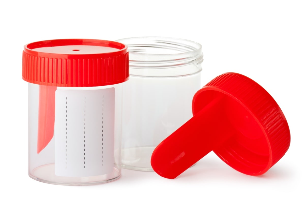
Small, empty plastic jars with lids that are used for urine samples.
A urine sample is collected in a special container and sent to a lab to help diagnose a UTI.

Lab tests

Urinalysis. A small amount of your child’s urine must be collected for this test. Babies and small children who are not toilet trained will have a small, thin, flexible tube called a catheter placed into the urethra to get a urine sample. This is needed because urine from collection bags, which can be taped around a baby’s diaper area, is often contaminated, or mixed, with germs and other substances found on the baby’s skin. If urine is contaminated, test results will not be accurate.

Parents may help preschoolers catch a clean urine sample in a special container, and older children and teens can do it by themselves.

A health care professional will look at the sample under a microscope for bacteria and white blood cells, which the body produces to fight infection. Bacteria also can be found in the urine of healthy children, so a bladder infection is diagnosed based on both your child’s symptoms and lab test results.

Urine culture. A health care professional must order a urine culture to find out what type of bacteria is causing your child’s infection. Lab workers will monitor how the bacteria multiply, usually over 1 to 3 days, to help determine the best treatment for your child.

Imaging tests

A health care professional may order imaging tests to find the cause of your child’s infection or to check for kidney damage.

Ultrasound. An ultrasound uses specialized sound waves to look at structures inside the body without exposing your child to radiation. During this painless test, your child lies on a padded table. A technician gently moves a wand called a transducer over your child’s belly and back. Ultrasound can create images of your child’s entire urinary tract. No anesthesia is needed.

Ultrasound may be recommended if your child

  • is younger than age 2 and has a bladder infection with a fever
  • has had repeated bladder infections at any age
  • has high blood pressure, poor growth, or a family history of kidney or bladder problems
  • doesn’t get better with treatment

An ultrasound may be scheduled right away or a few weeks or months after your child’s illness has passed.

An ultrasound exam on a child. A technician passes a wand over a child’s lower back, which sends an image to a computer screen.
An ultrasound produces images of a child’s kidneys, ureters, bladder, and urethra. The test can help find the cause of a bladder infection.

Voiding cystourethrogram (VCUG). A voiding cystourethrogram uses x-rays of the bladder and urethra to show how urine flows. A catheter is used to fill your child’s bladder with a special dye. Then x-ray pictures are taken before and after your child urinates. A VCUG can show if urine flows backward from the bladder into the ureters or kidneys, a condition called vesicoureteral reflux (VUR). Anesthesia is not needed for this test, but your child may be offered a calming medicine, called a sedative.

Read more about imaging tests of the urinary tract.

Last Reviewed April 2017
Share this page
Facebook X Email WhatsApp LinkedIn Reddit Pinterest

This content is provided as a service of the National Institute of Diabetes and Digestive and Kidney Diseases (NIDDK), part of the National Institutes of Health. NIDDK translates and disseminates research findings to increase knowledge and understanding about health and disease among patients, health professionals, and the public. Content produced by NIDDK is carefully reviewed by NIDDK scientists and other experts.

The NIDDK would like to thank:
Saul P. Greenfield, MD, FAAP, FACS, State University of New York at Buffalo School of Medicine; Jeffrey M. Saland, MD, MSCR, Icahn School of Medicine at Mt. Sinai